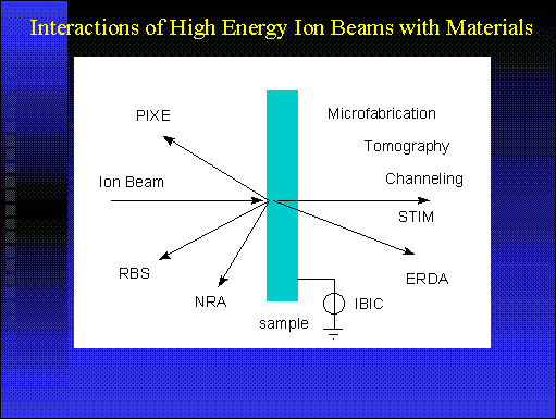
When a fast moving charged particle collides with an atom there is a
reasonable probability that an electron will be ejected from an inner
atomic shell. Subsequently, the electrons in the oter shells re-arrange
themselves with the emission of a quantum of energy (X-ray), the energy
of which is characteristic of the parent atom. The measurement of these
characteristic X-rays enables the chemical composition of a sample to
be determined with high quantitative accuracy and sensitivity (10-20
ppm for Na to Cl
, and 1-10 ppm for Ca and higher in the periodic table).
More details about RBS
As the ion traverses the sample, there is a small probability that a
direct elastic nuclear collision will also occur causing the ion to
recoil out of the sample. By masuring the energy of recoiling ions,
information can be obtained on the concentration a
nd depth distribution of major constituents of the sample, such as the
light element matrix C, N and O. RBS is complementary to PIXE and allows the sample matrix to be
characterised, thus making quantitative measurements of the trace e
lements more accurate.
More details about STIM
For most biological samples (tissue sections, isolated cells), the high
energy particle beam passes through the sample, loosing energy by
collisions with electrons. By measuring the energy loss of individual
particles in the transmitted beam, information on the density or
thickness of the specimen can be obtained.
NRA
Nuclear Reaction Analysis: Measurement of reaction products such as
gamma rays, alpha particles and protons following nuclear reactions
between the incident ion and the target atoms. Used mainly for light
ions up to Mg in the periodic table and thier isot
opes.
ERDA
Elastic Recoil Detection Analysis: Measurement of recoiling atoms
following elastic nuclear collisions at a glancing angle. Used mainly
for profiles of very light ions such as hydrogen or deuterium.
IBIC
Ion Beam Induced Current is a technique which images the active regions
in micro-electronic devices, by measuring the induced current caused by
a penetrating beam of focused ions. This technique enables active
regions to be assessed on-line for resistance to radiation.
Channeling
If the direction of the ion beam is aligned with a plane or axis of a
crystal, then channeling can occur and the ions travel through the
crystal with much reduced energy loss. This phenomenon can be used to
measure crystal quality and the mapping of the l
attice plane directions (strain measurements) when used in conjunction
with PIXE and RBS. When used
with STIM, the technique can provide information
on faults such as lattice distortion and disloc
ations at depth in the crystal.
Ion Microbeam Tomography
The use of a penetrating focused beam of high energy ions to produce
information at depth on the elemental constituents and internal
structure of a specimen. By scanning the beam over the specimen at many
orientations, 3D information can be obtained and 3
D images generated.
Ion Microbeam Microfabrication
The use of finely focused and penetrating high energy ion beams to form
deep (~10 µm) and narrow (~100 nm) channels in resists used in
lithography.